Introduction
In this article we will focus on the benefits of performing an Chest X Ray . Find out details about what an X Ray does, how a doctor or a Radiologist diagnoses your illness and how it help to ease your concerns.
Table of Contents
- Introduction
- Medical uses of Chest X Ray
- How good is Chest X Ray for diagnosis?
- A few diseases that are seen in Chest X Ray
- Further Reading
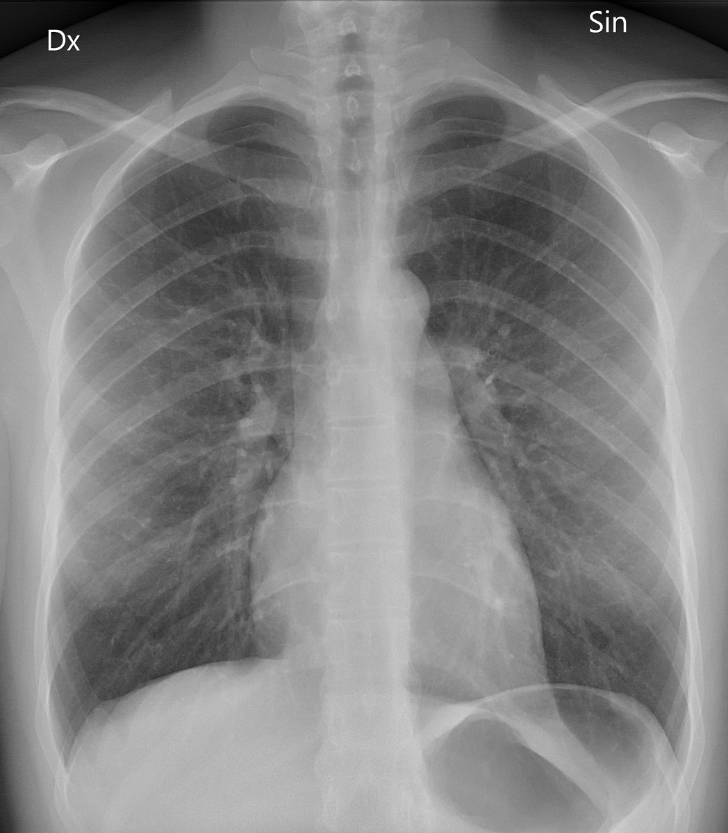
A Chest X Ray is a vital tool in the hands of a doctor. It is very useful to find out the appearance of the chest, to determine if there might be any diseases lurking inside.
X Rays are taken by exposing the person in concern to a small amount of ionizing radiation of 0.02 mSv.
One X Ray corresponds to a background radiation equivalent time of about 10 days
https://en.wikipedia.org/wiki/Chest_radiograph
Medical uses of Chest X Ray
A Chest X Ray becomes a vital tool to find out underlying diseases such as:
- Air in the Lungs – Pneumothorax
- Pneumonia (Inflammation of the air sacs in the lungs)
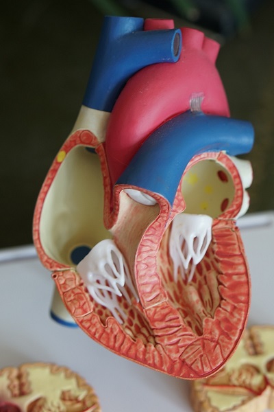
- Heart related concerns
- Fractures of the ribs
- Hiatus Hernia (Herniation of the stomach walls into the Esophagus)
Apart from the above the chest X Ray can be useful to determine if the person may be having ground glass appearance, which may further be determined with an HRCT, and also rule out Tuberculosis affection.

How good is Chest X Ray for diagnosis?
There was a time where there were not may tools available for diagnosis apart from an Chest X Ray. While a few other places had MRIs and CT Scans; the cost of these investigations was such that they were beyond the reach of the ordinary. Then there were simple Chest X Rays which did not have good quality imaging. They were so much clouded, that it was with high skill that the Radiologist would read the same! Later computers brought on more improvement, making the X Rays more better.
Since early 2000s, computer generated and AI guided Chest X Rays are well prepared, reducing the amount of Radiation exposure as well.

A few diseases that are seen in Chest X Ray
Nodules
One may be able to see nodules. Nodules are small round shape appearances on the film. These may be due to Tuberculosis, infection, reduced blood flow or even sometimes due to cancers. So when you see a nodule, do not think of Cancer first, rule out other possibilities.
Cavities
Cavities are seen as hollow structures in the lungs. These may be due to thickening of the pleura wall, infections or an infarct.
Diffuse shadowing
This type of appearance on the filmrecently gained a lot of popularity owing to the COVID 19 infection. One may look for crisscross lines, rings, cysts, ground glass appearance or consolidations on the Chest X Ray.
The location is more important in this case. Depending on the location, the radiologist may pin point on the probable disease cause.
Cystic appearance
Cystic appearances in the Chest X Ray may point towards Alveolitis. Alveolitis gives a honey comb appearance in the lung. Cystic appearance can also come in the Bronchi of the lung.
Thus it can be noted that the filmis a great tool to find out the approximate cause of disease and may further help in finding out which medication may be required to help the patient in improving in overall health standards.
Consolidation appearances
Consolidation refers to reorganization and improvement in the pertinent lung disease. These can be seen in diseases like pneumonia, alveolar hemorrhage, vasculitis and alveolar cell carcinoma.
Acute Respiratory Distress Syndrome
ARDS can be seen with a complete white appearance on the report. This happens when the disease spreads very fast and is observed all over the lungs. The person experiences shortness of breath, rapid breathing and bluish skin appearance.
Further Reading
To know more about Wellness and how it can be connected to Chest X Ray, check out our video on Wellness in Hindi Language. Also check Tamhane Clinic website for further articles on X Rays to gain a deeper understanding.
Want to buy a few items related to X Rays? Check our shop over here.